
|
2015 Biology |
||||
|
The analysis of lasso peptides by circular dichroism Gouffran Célia |
||||
|
Introduction Lasso peptides constitute an interesting and original class of bacterial peptides. They are composed of a macrolactam cycle in which the C-terminal tail is trapped by cumbersome residues or disulfide bridges. The lasso peptides structure is all the more interesting that lasso peptides do not have all the same spectroscopic signature. Experimental conditions Different lasso peptides of type 2 (Mcc J25 and Capistruin) and 3 (BI-32169) were analyzed by circular dichroism at synchrotron SOLEIL, using a 0.02 cm pathlength cistern. The lasso peptides were taken into phosphate buffer at 10 mM and pH 7. The experience was realized at 25°C. Results The three CD spectra do not have the same appearance. They differ by the presence or not of the 230 nm band. In view of their amino acid sequence, the aromatic amino acids like tryptophan (W), tyrosine (Y) and phenylalanine (F) may be involved on this 230 nm band. Conclusion The circular dichroism enables to characterize the spectroscopic signature of lasso peptides. It gives some information about the presence of the 230 nm band which is caused by the interaction between several aromatic amino acids. Moreover, the kinetic of the lasso structure loss can be followed by circular dichroism. |
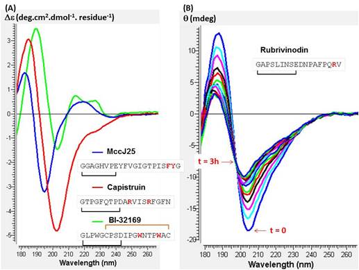 CD spectra of Mcc J25 (blue), Capistruin (red) and BI-32169 (green) at 25°C (A) and the denaturation of Rubrivinodin at 90°C for 3 hours (B) ; 0.02 cm pathlenght cistern ; phosphate buffer 10 mM pH7 |
|||
|
National Musuem of Natural History |
|
|||
ANALYSE & CONTROLE, le MASTER Batiment Bertholet - 22 avenue Gaston Berger 69622 Villeurbanne Phone : 04-72-44-79-88 mail : master-analyse-controle@univ-lyon1.fr |
DIRECTOR Jerome RANDON SECRETARY |
TECHNICAL STAFF Herve DELEPINE Julie BERTRAND Didier FOURNIER |
EDITORIAL OFFICE Editorial Director : Jerome RANDON webmastering : Chahira YAHIAOUI |