
|
2015 Instrumentation |
||||
|
Gold nanoparticle physicochemical characterization Usanase Gisèle |
||||
|
Introduction Gold nanoparticle (AuNP) are used in various applications covering electronics, biosensors, biomedical imaging and in vitro biomedical diagnosis due to its strong interaction with light. Indeed AuNP have the ability to absorb and scatter visible light depending upon their size, shape and agglomeration state. AuNP must be well characterized not only under dried state but mainly in dispersed media. The characterization step is essential in the development of nanoparticles; because their sizes, size distribution, and morphology influence their physicochemical properties. Experimental conditions The hydrodynamic diameter of AuNP was determined by DLS using the Zetasizer from Malvern Instrument at room temperature (25 C°). For TEM a small drop of suspension was deposited on a microscope grid (copper support covered with carbon) and slowly dried in open air. The dry samples were observed by TEM with a Philips CM120 microscope under 120 kV acceleration voltage. Absorbance was examined using a Spectrophotometer from Shimadzu. The maximum wavelength (λmax) and FWHM (Full Width at Half Maximum) were extracted from the obtained spectrum then used to estimate the size using a standard curve and to calculate size particle using the Mie-Lorenz theory. Results Under TEM observations, the AuNP had spherical shapes (Fig. 1) with an average particle size smaller than 30 nm. TEM Images showed aggregation between some particles for F7a, F7b and F2 (Fig.1). However fairly detached and very homogeneous particle were found F6a, F6b, F3, and F5 (Fig 1) with narrow size distribution. Conclusion The AuNP were prepared successfully and their particle size, size distribution and morphology were verified through different methods. Furthermore, with this study we now have all information needed to choose the right AuNP to use in biomedical imaging techniques. |
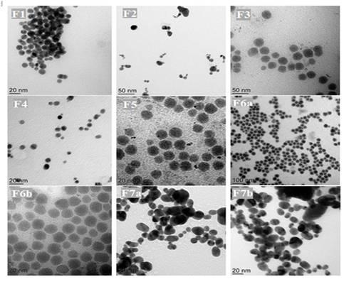 TEM micrograph of colloidal dispersion AuNP: F1, F2, F3, F4, F5, F6 (a, b), F7 (a, b) 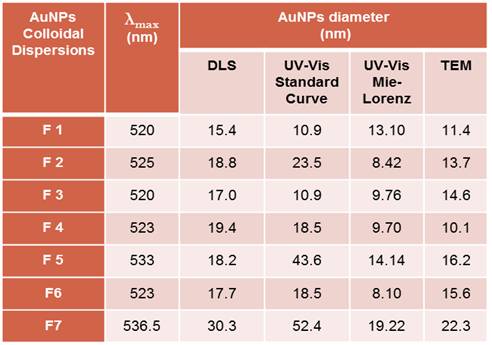 AuNP diameter (nm) of each dispersion obtained by all analytical technics used |
|||
|
LAGEP |
|
|||
ANALYSE & CONTROLE, le MASTER Batiment Bertholet - 22 avenue Gaston Berger 69622 Villeurbanne Phone : 04-72-44-79-88 mail : master-analyse-controle@univ-lyon1.fr |
DIRECTOR Jerome RANDON SECRETARY |
TECHNICAL STAFF Herve DELEPINE Julie BERTRAND Didier FOURNIER |
EDITORIAL OFFICE Editorial Director : Jerome RANDON webmastering : Chahira YAHIAOUI |