
|
2017 Biology |
||||
|
Determination of the type of glycosylation by SDS-PAGE Uteem Imteyaz |
||||
|
Introduction This study in the research unit UMR 7245 of the CNRS and the National Museum of Natural History is the result of a collaboration with the CEA of Cadarache. Experimental conditions In order to know the type of glycosylation, we used two methods of deglycosylation, one specific for N-glycosylation (enzyme reaction by N-glycosidase F) and one specific for O-glycosylation (chemical reaction of β- elimination). Results Regarding N-glycosylation, by observing 806+ (treated with the kit) versus 806- (untreated), no difference was observed. Similarly for 807+ and 807-. It is deduced that the bacteria are not N-glycosylated (figure 1). As regards O-glycosylation, for the 806+ and 806- proteins, no task was observed because these samples could not be placed at a pH between 6 and 8. In parallel, it is noted that the spots of the proteins 807+ compared to the 807- are less marked (figure 2). We can deduce that our strains are probably O-glycosylated, and we can not confirm it because we do not have control samples. Conclusion The bacterial strains 806 and 807 are not N-glycosylated. On the other hand, strain 807 is probably (because there is no control sample) O-glycosylated. |
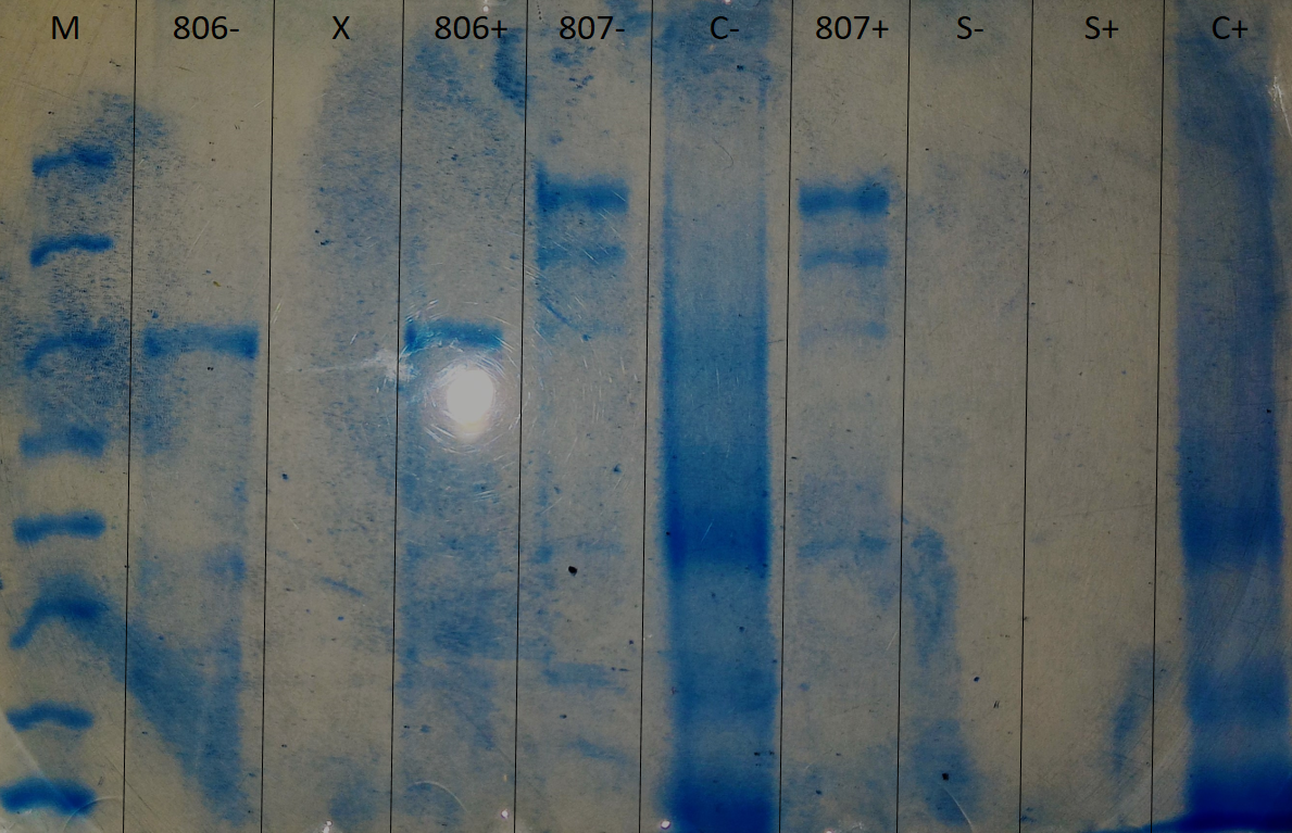 SDS-Page 6% polyacrylamide electrophoresis, executed to determine if the glycosylation is of the N-glycosylation type. 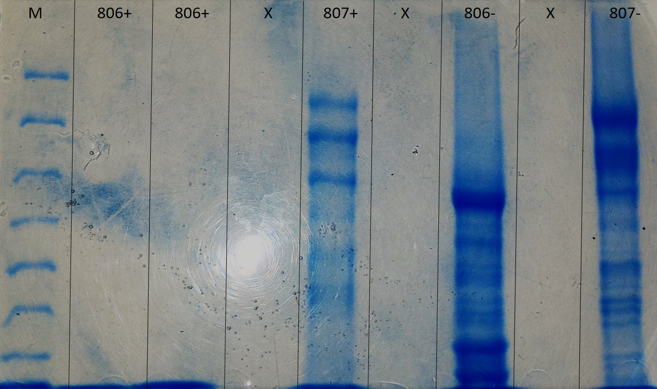 SDS-Page 6% polyacrylamide electrophoresis, executed to determine if the glycosylation is of the O-glycosylation type. |
|||
|
Muséum National d'Histoire Naturelle, UMR 7245 CNRS/MNHN |
|
|||
ANALYSE & CONTROLE, le MASTER Batiment Bertholet - 22 avenue Gaston Berger 69622 Villeurbanne Phone : 04-72-44-79-88 mail : master-analyse-controle@univ-lyon1.fr |
DIRECTOR Jerome RANDON SECRETARY |
TECHNICAL STAFF Herve DELEPINE Julie BERTRAND Didier FOURNIER |
EDITORIAL OFFICE Editorial Director : Jerome RANDON webmastering : Chahira YAHIAOUI |