|
Introduction
Botrytis cinerea is a necrotrophic fungus, which affects many plant species, especially bunches of grapes. New fungicides have been searched to control it. On the one hand there are fungicides acting on fungi respiratory chain, on the other hand on sphingolipids biosynthesis, which are components of cell membrane. To find a simple way to elucidate Mode Of Action, any methods to analyze the modifications in sphingolipids biosynthesis induced by fungicides are welcome.
Compounds with long hydrocarbon chains as sphingolipids and their precursors can be analyzed by LC-MS/MS , GC-MS but also by HPTLC. A historical technique as the thin layer chromatography presents many advantages compared to many others. Sphingolipids cannot be analyzed with GC-MS without a prior derivatization to give them volatility. With LC-MS/MS, their fat nature will clog the apparatus involving a regular maintenance. In comparison, HPTLC allows the analysis of at least twenty samples in the same short time and is intended to free itself of the sample preparation.
This work is about developing a method that allows separation and visualization of sphingolipids on the plate by HPTLC.
Experimental conditions
As first intention, standards analyzed are 3-Ketodihydrosphingosine (Matreya LLC), dihydrosphingosine, phytosphingosine (Sigma Aldrich).
The first assays carried out with CHCl3/MeOH 2/1 as a mixture to dissolute compounds with CHCl3/MeOH/NH4OH 65/35/5 v/v/v as a mixture for migration, which stems from literature (Weiss & Stiller, 1972).
Finally, the second assays presented were done with DCM/MeOH 2/1 v/v as new mix to dissolute compounds and DCM/MeOH/H2O 50/40/10 v/v/v as mixture for migration.
The compounds were at the rate of 1 mg/ml in the dissolution mixture.
Standards applied at different volumes (2; 5; 10; 20 µl depending to the assay) on a silica gel 20x10 cm plate (Merck) with ATS4 (Camag) and migrated in a vertical glass chamber (Camag).
The visualization of spots with ninhydrin were made by immersion of the plate in the reagent during 4 seconds followed by heating the plate for 5 minutes at 110°C with a heating plate (Camag). Ninhydrin was used for its sensitivity linked to the reaction with primary amines.
Results
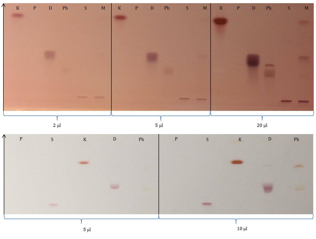
By changing the elution mixture, selectivity of migration has been modified. First plate (Fig. 1A) shows spots, which spread on the plate and having drag. Using ternary mixture allows 3-Ketodihydrosphingosine to migrate to the front while serine does not migrate. Moreover, the ammonia used in the migration mixture reacted with ninhydrin, coloring the plate in red.
In the second case, changing chloroform by dichloromethane allowed the decrease to the spreading standards. Changing ammonia by water allowed a white background plate instead of a red one.
The dyeing reagent showed its limit. Despite its specificity, the reagent lacks sensitivity and complementary tests had shown that 1 µg spread on the plate was hardly visible and that 0,1 µg was invisible for example for 3-Ketodihydrosphingosine.
Conclusion
Although improvement are still required, preliminary results seems to demonstrate that HPTLC could be an alternative to other method for analyzing easily fungal sphingolipids.
A gradient mode analysis will allow focalization of spots and the decrease of spread. A coupling with mass spectrometry will offer a better sensitivity and an easier analysis in a complex matrix.
|
|

Figure 1 - Sphingolipids precursors migration and visualization with ninhydrin
A: standards separation with CHCl3/MeOH/NH4OH, applied about 2; 5; 20 µl (left to right).
B: standards separation with DCM/MeOH/H2O, applied about 5; 10 µl (left to right).
Legend:
• K: 3-Ketodihydrosphingosine
• P: Palmitoyl CoA
• S: L-Serine
• D: Dihydrosphingosine
• Ph: Phytosphingosine
• M: Mix
|


